Anemia in Dogs — Low HCT and RBC Results
Wondering what a full interpretation looks like? See a sample report →
What a Low HCT and RBC Actually Confirm
The hematocrit, often written HCT and closely tracked by the older term PCV, is simply the fraction of the blood made up of red cells once they are packed together. The RBC count is the number of those cells. When both fall below the reference interval, the panel is telling your veterinarian one clear thing: this is anemia, meaning the blood is carrying less oxygen than it should. That is the whole of what the two figures confirm. They do not say whether the dog is losing red cells, destroying them, or failing to make enough of them, and they do not say how the dog is coping. Those answers come from the reticulocyte count, the red-cell indices and the rest of the panel, and from the dog standing in front of the veterinarian. A full CBC and chemistry panel is the natural setting for reading these values, and the step-by-step approach to a complete dog report shows where red-cell numbers sit among everything else.
It is worth being clear about what the two figures are not. They are not a measure of how much blood the dog has: a dog that has just bled heavily can still show a near-normal hematocrit for the first hours, because what has been lost is whole blood – cells and fluid together – and the proportion between them has not yet changed. It is only as the body pulls fluid back into the circulation, or as fluids are given, that the dilution becomes visible and the number falls to reflect the loss. The reverse is also true: a dehydrated dog concentrates its blood, so an anemia can hide behind a hematocrit that looks acceptable until rehydration unmasks it. This is why your veterinarian reads the red-cell values against the dog’s hydration and the clinical picture, and why a single value taken at the wrong moment is repeated rather than trusted.
HCT, RBC and Hemoglobin – Reading Them as a Set
HCT and RBC move together in anemia, which is why a veterinarian reads them as a pair rather than fixating on one. Hemoglobin, the oxygen-carrying pigment inside each cell, usually falls in step with them and is the value that most directly explains why an anemic dog tires easily. When the three do not agree neatly, the red-cell indices step in to explain the discrepancy: the average size of each cell and how much hemoglobin it holds tell the veterinarian whether the cells are normal, small and pale, or large and immature. Those indices are interpreted in their own right – the MCHC reading and questions about what a high MCH means are read alongside the headline anemia. Seen as a set, these numbers begin to describe the kind of anemia, not just its presence. The mirror-image situation, when these same values run high, is covered in high hematocrit and high RBC in dogs.
Where the Normal Line Sits
Reference intervals shift between laboratories with the analyser and method used, so the range printed on your own dog’s report is always the final word – and breed and age move the line as well, which is taken further for the bigger breeds in anemia in large-breed dogs. As an evidence-based guide, the verified canine red-cell intervals are below. The reticulocyte count is listed with them because it, more than any single red-cell figure, decides which direction the investigation takes.
| Parameter | Normal reference range (dog) |
|---|---|
| HCT (hematocrit) | 41-58% |
| RBC (red blood cell count) | 5.7-8.5 x10^6/uL |
| Hemoglobin (HGB) | 14.1-20.1 g/dL |
| MCV (mean cell volume) | 64-76 fL |
| MCHC | 33-36 g/dL |
| Reticulocytes | 0.2-1.5% |
See the full reference intervals for the dog — every value cited to the laboratory document or paper it came from.
A value a little below the interval in a bright, well dog and the same value in a collapsed one are not the same finding, which is precisely why no red-cell number is read in isolation.
Regenerative or Not – The Question That Splits the Workup
Once anemia is confirmed, the single most important branch point is whether the bone marrow is responding. The reticulocyte count answers it. When reticulocytes rise, the marrow has sensed the loss and is pushing out young cells to replace them – a regenerative anemia, which usually means red cells are being lost through bleeding or destroyed faster than normal. When reticulocytes stay flat despite a low HCT, the marrow is not keeping up; the anemia is non-regenerative, and the investigation turns toward the marrow itself or the chronic diseases that suppress it. This split shapes everything that follows, and it maps roughly onto the broad categories below.
| Marrow response | What it suggests | Typical next direction |
|---|---|---|
| Regenerative (reticulocytes up) | Red cells being lost or destroyed | Look for bleeding or hemolysis |
| Non-regenerative (reticulocytes low) | Marrow not replacing cells | Investigate chronic disease or marrow |
| Early or borderline | Response may not show yet | Recheck and reassess the trend |
Where destruction is the suspect, supporting clues often appear elsewhere on the panel – a rise in bilirubin can point toward hemolysis, and a markedly high total bilirubin sharpens that question further. Because immune-mediated destruction can take platelets down with the red cells, a paired drop in platelets is something your veterinarian watches for too.
Where the Red Cells Are Going
Once the marrow question is settled, the search narrows to a route. Blood loss is the most immediately treatable of the three, and in dogs it is often hidden rather than dramatic: bleeding into the stomach or intestine from ulceration, from a tumour, or as a consequence of anti-inflammatory medication; slow loss into the abdomen from a ruptured splenic mass; or, in puppies and heavily parasitised dogs, a burden of fleas or intestinal worms large enough to matter. What these share is that the dog loses both red cells and iron over time, so the cells that are made can end up smaller and paler than they should be.
Destruction is a different picture. The red cells are produced and then broken down early, which is why bilirubin can rise and why the gums or the whites of the eyes may take on a yellow tinge. Immune-mediated destruction is the best known form in dogs and can appear on its own or alongside another illness, an infection or a recent drug. Other causes are mechanical or toxic: cells damaged as they pass through abnormal vessels or a tumour, damaged by substances such as onion, garlic or zinc from a swallowed coin, or parasitised directly by tick-borne organisms. These usually behave as regenerative anemias once the marrow has had a few days to answer.
The third route is under-production, and it tends to be slow and quiet. Chronic kidney disease is the classic example, because a damaged kidney makes less of the hormone that tells the marrow to produce red cells; the anemia then tracks the kidney disease rather than any bleeding. Long-running inflammation, infection or cancer can blunt production in a similar way, and disorders of the marrow itself, along with a small number of drugs, can suppress it directly. The practical consequence is that a non-regenerative anemia sends your veterinarian back to the rest of the panel and to the dog’s history rather than hunting for a bleed.
| Route | Clues that fit it | Where the search goes |
|---|---|---|
| Loss | Regenerative response; black or tarry stools; recent surgery, trauma or anti-inflammatory treatment; heavy parasite burden; smaller, paler cells if it has run for weeks. | Check the stool for hidden blood, image the abdomen, review medication and parasite control. |
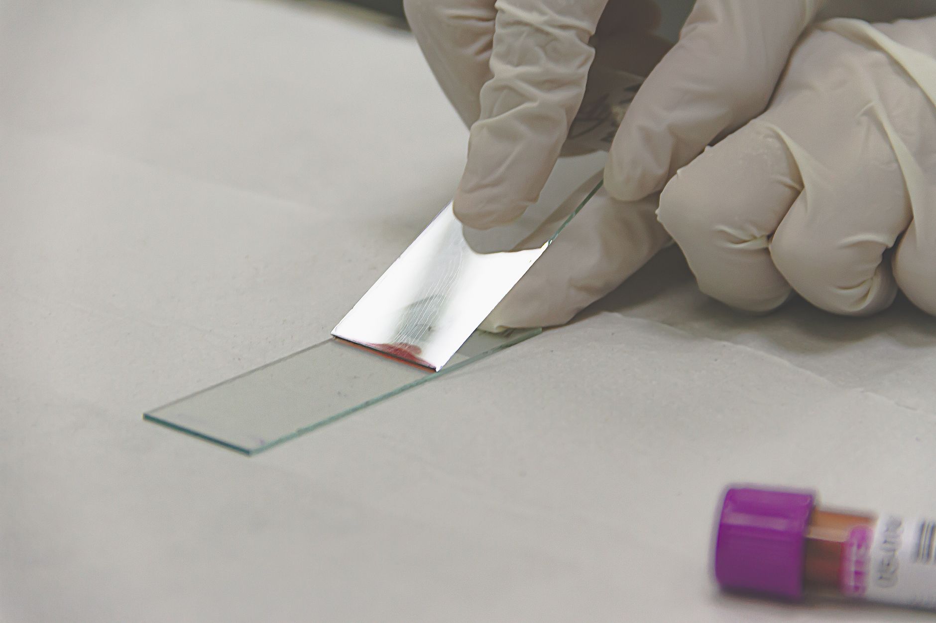
| Destruction | Regenerative response with rising bilirubin; discoloured urine; red cells clumping or looking small and dense on the smear; platelets sometimes falling with them. | Read the smear, check bilirubin, look for an infection, a toxin or a recent drug behind it. |
| Under-production | Flat reticulocytes; kidney markers or inflammatory changes elsewhere on the panel; a long, slow history rather than a sudden collapse. | Assess kidney function and chronic disease first; sample the marrow only when those do not explain it. |
How Fast It Happened Changes Everything

Two dogs can arrive with the same hematocrit and be in completely different trouble. A dog whose red cells fell over months has had time to adapt: the heart works harder, the tissues take more oxygen from each cell, and the dog may still be walking, eating and behaving almost normally. A dog that has bled into its abdomen this morning has had no such time. That is why your veterinarian weighs the history and the examination as heavily as the number, and why a value that is merely monitored in one dog is an emergency in another.
The signs that carry the most weight are the ones saying the tissues are short of oxygen now: very pale or white gums, weakness or collapse, breathing quickly or with effort at rest, a racing heart, or a dog that is cold and dull. A yellow tinge to the gums or skin adds the suspicion of destruction, and a distended, painful abdomen raises the possibility of bleeding inside. Any of these belongs in a consulting room the same day rather than at the next routine appointment.
Two practical questions come up once the dog is stable. The first is whether the anemia is likely to return: a bleed that was stopped, a drug that was withdrawn or a parasite burden that was treated usually does not come back if the cause stays fixed, whereas an immune-mediated process can relapse as treatment is reduced, which is why those dogs are rechecked on a schedule rather than discharged. The second is what the owner should watch for at home between visits. Gum colour, breathing at rest, appetite and stamina on a normal walk are the four things that change earliest, and a dog that slips backwards on any of them deserves an earlier recheck than the one in the diary. Written down together with the dates of each blood test, those observations often tell your veterinarian more about the direction of travel than a single repeat sample would on its own.
Stabilisation, when it is needed, is decided by how the dog is coping rather than by a threshold on the report. Some dogs need only the cause addressed – a bleeding source controlled, a drug stopped, treatment started for an immune-mediated process. Others need support first, which may include a transfusion, and that decision belongs to the veterinarian examining the dog. Once the immediate risk has passed, the red-cell picture is rechecked on a schedule that matches the cause, because what matters then is the direction of travel rather than any single reading.
| What you notice at home | Why it changes the urgency |
|---|---|
| Very pale or white |