High GGT and High ALP in Cats – What the Pattern Means
Wondering what a full interpretation looks like? See a sample report →
Two Enzymes That Tell the Same Story
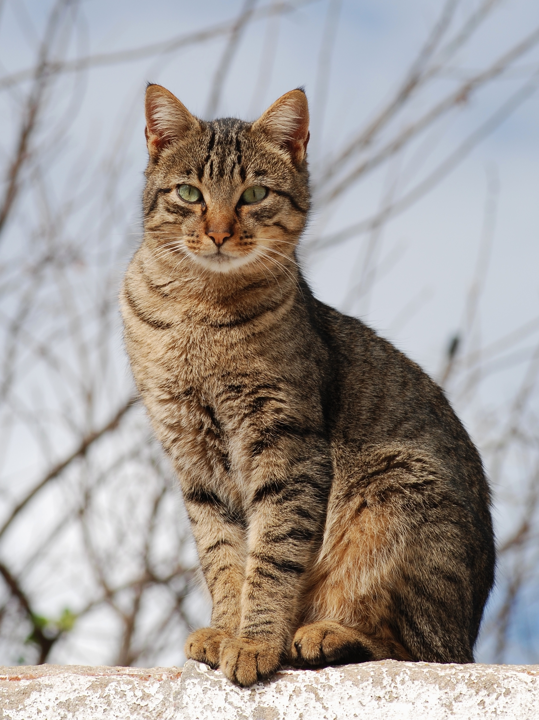
Liver enzymes fall into two broad camps, and understanding which camp GGT and ALP belong to is the whole key to reading this pattern. Some enzymes – alanine aminotransferase and aspartate aminotransferase – live inside liver cells and spill out when those cells are injured. They are leakage markers. Gamma-glutamyl transferase and alkaline phosphatase are different. They are cholestatic enzymes: their activity rises when the movement of bile through the liver and bile ducts is impaired. When both of them are elevated at once, they are telling the same story from two angles, and that story is about bile flow.
This is the single most important correction to make about this pattern. A combined GGT and ALP rise is not a measure of how badly liver cells are dying; it is a signal that bile is not draining as it should. That distinction changes what the veterinarian looks for next and which structures get imaged. It is also why these two enzymes are so often read together with the leakage markers covered in our guide to high ALT and high AST in cats – the two patterns answer different questions.
There is one feature of feline biochemistry that makes this pair especially worth paying attention to. In cats, ALP has a notably short half-life, far shorter than in dogs. The practical effect is that the enzyme clears quickly, so the body has to be actively producing more of it for the value to stay raised. A cat does not carry a lingering, meaningless ALP elevation the way a dog sometimes can. That is why even a modest rise tends to be taken seriously rather than waved away as a quirk of an ageing animal.
It also means the old shorthand that “ALP just reflects bone growth” does not apply to an adult cat the way it might to a young, growing dog. In a mature cat, a persistently raised ALP is far more likely to be telling you about cholestasis – or about a fatty liver – than about the skeleton. Reading the value through a feline lens, rather than a borrowed canine one, matters. Our overview of feline-specific ALP thresholds expands on why the cat is its own case.
What Each Enzyme Is Actually Reporting
It helps to take the two enzymes one at a time before reading them together, because each one narrows the search in its own way.
GGT sits in the cells lining the bile ducts and the canalicular surface of liver cells. When bile flow is obstructed or the bile ducts are inflamed, GGT production climbs and the blood level rises. It is a fairly specific signpost: a raised GGT pushes the veterinarian toward the biliary tract rather than toward something vague. It does not indicate that liver cells are leaking, and it is not a measure of how many cells have been damaged. Our dedicated piece on evaluating biliary disease through GGT in cats goes deeper on this single marker.
ALP is induced when bile flow is impaired, so it rises with cholestasis as well. In the cat, because of that short half-life, the enzyme is a sensitive early reporter – it moves when something is genuinely going on. The trade-off is that ALP is less specific than GGT about exactly where the trouble is, which is precisely why the two are most powerful when read as a pair rather than alone. A fuller treatment of how the cholestatic enzymes sit within the whole liver picture is in our interpretation guide to ALT, AST, ALP and GGT in cats.
When you put them side by side, a consistent rise in both points firmly at cholestasis. A divergence between them – one clearly up, the other lagging – is itself a clue, and that divergence is where some of the most useful distinctions in feline liver disease are made.
Reading the Direction the Enzymes Point
The combined pattern is a starting point, but the relationship between the two enzymes refines it. Because GGT is the more specific biliary marker and ALP is the more sensitive one, the way they move relative to each other narrows the field of likely causes. None of this confirms a diagnosis – it shapes the next set of questions and tests.
The most clinically useful contrast in cats is the one between cholangitis and hepatic lipidosis. Inflammation of the bile ducts – cholangitis, often part of the broader picture explored in our article on GGT as a cholangitis marker in cats – tends to lift both GGT and ALP together, with GGT often standing out. A fatty liver behaves differently: ALP is frequently the more prominent rise while GGT stays normal or climbs only slightly. So a cat with both enzymes clearly elevated is, if anything, leaning toward biliary inflammation rather than a textbook fatty liver – although the two conditions can coexist, and only the full workup separates them. The way these conditions overlap is laid out in our piece on distinguishing cholangitis, hepatic lipidosis and cancer in cats.
The table below sketches how a veterinarian thinks through the pattern. Read it as a set of leanings, not as a lookup that hands you an answer – the cat’s appetite, weight, jaundice and the rest of the panel always carry more weight than any single combination of enzymes.
| Pattern | What it tends to suggest | How the vet follows up |
|---|---|---|
| Both GGT and ALP elevated together | Cholestasis with biliary-tract involvement – inflammation of the bile ducts is a leading consideration. | Abdominal ultrasound of the liver and bile ducts, bilirubin and bile acids, and pancreatic markers given how often the two are linked in cats. |
| ALP up, GGT normal or only mildly raised | A pattern that leans toward hepatic lipidosis rather than primary bile-duct disease. | Assessment of appetite and recent weight loss, ultrasound of the liver, and prompt attention if the cat is not eating. |
| Cholestatic enzymes up alongside raised ALT and AST | Bile-flow trouble accompanied by liver-cell injury – a mixed hepatobiliary picture. | Read together with the leakage markers and bilirubin to judge how much of each process is present. |
| Cholestatic enzymes up with raised pancreatic markers | Pancreatic disease disturbing nearby bile flow – common in the feline abdomen. | Pancreatic-specific testing and imaging of the pancreas as well as the liver. |
See the full reference intervals for the cat — every value cited to the laboratory document or paper it came from.
Where the Reference Intervals Sit
Reference intervals depend on the analyser and the method, so the range printed on your own cat’s report is always the final word. As an evidence-based guide, the verified feline intervals for the cholestatic enzymes – and for the leakage markers and bilirubin that frame them – are listed below. Notice how narrow the feline GGT interval is: in a healthy cat the value barely registers, which is part of why even a small rise is meaningful.
| Parameter | What it reports | Normal reference range (cat) |
|---|---|---|
| GGT | Cholestatic marker; biliary-tract involvement | 0-2 U/L |
| ALP | Cholestatic, inducible enzyme; short half-life in cats | 11-49 U/L |
| ALT | Hepatocellular leakage marker | 28-109 U/L |
| AST | Leakage marker; liver and muscle | 17-48 U/L |
| Total bilirubin | Pigment cleared by the liver; rises in cholestasis or haemolysis | 0-0.1 mg/dL |
The point of showing these together is context. GGT and ALP describe bile flow; ALT and AST describe cell injury; bilirubin describes whether the liver is keeping up with its job of clearing pigment. A cat whose cholestatic enzymes are above the interval but whose bilirubin is still within range may be at an earlier stage than one in whom bilirubin has begun to climb. For a wider tour of how the chemistry and the blood count fit together, our complete cat blood panel reading guide is a good companion.
What Sits Behind the Pattern in Cats
Several feline conditions converge on a cholestatic enzyme rise, and they are worth knowing not so you can self-diagnose but so the workup makes sense when your veterinarian orders it. The list below describes the usual suspects, not a ranking of likelihood for any individual cat.
- Inflammation of the bile ducts (cholangitis). One of the more common feline liver diseases, it directly disturbs bile flow and tends to raise both GGT and ALP. It frequently overlaps with intestinal and pancreatic inflammation in cats, which is why the abdomen is assessed as a whole.
- Hepatic lipidosis (fatty liver). When a cat stops eating, fat mobilises into the liver and overwhelms it. ALP is typically the louder enzyme here, with GGT often staying quieter – a contrast that helps separate it from bile-duct disease. The condition is covered in depth in our article on hepatic lipidosis lab findings and emergency nutrition.
- Pancreatic disease. Because the feline pancreatic and bile ducts sit so close together, inflammation in the pancreas can impede bile flow and lift the cholestatic enzymes. This anatomical closeness is a recurring theme in cat medicine.
- Obstruction of the bile ducts. A physical blockage – by a thickened gallbladder, a mucus plug or a mass – backs bile up and drives the cholestatic enzymes upward, often with bilirubin following.
What unites these is the mechanism, not a single named disease. The pattern says bile is not flowing freely; finding out why is the job of imaging, the rest of the panel, and sometimes sampling of the liver itself. A combined enzyme rise should always be read alongside the leakage markers, because a mixed picture – cholestasis plus cell injury – is common, and the balance between the two shapes the plan. Our guide to decoding elevated liver enzymes in cats walks through how these threads are woven together.
How Far the Enzymes Take You – and Where Other Tests Begin
GGT and ALP tell the veterinarian that bile flow is disturbed. What they cannot do is measure whether the liver is still doing its work, and that gap is filled by other tests. Bilirubin and bile acids assess function rather than enzyme activity – they show whether the liver is keeping up with clearing pigment and recycling bile. A cat can have raised cholestatic enzymes while function is still holding, or it can have both abnormal at once, and that difference changes how urgent the situation is.
Imaging is the natural next step. An abdominal ultrasound looks directly at the liver, the gallbladder, the bile ducts and the pancreas, and it often answers the question the enzymes only raised. In cats, because liver, pancreatic and intestinal disease so often travel together, the scan rarely stops at the liver. From there, sampling of the liver may be considered when the picture remains unclear – but that is a decision your veterinarian makes based on the whole animal, not on the enzyme values alone.
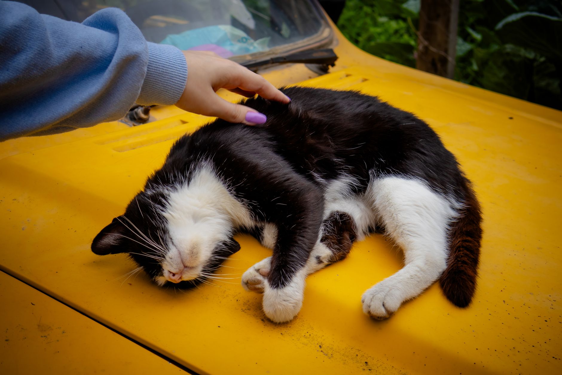
On the question of breed and age that the image above raises: there is no strong breed predisposition for a cholestatic enzyme pattern in cats, and individual history matters far more than pedigree. Age is relevant chiefly because bile-duct disease and fatty liver are seen more often in middle-aged and senior cats, and because the meaning of an ALP rise in an adult cat is not softened by any “still growing” allowance the way it might be in a young animal. The number deserves the same scrutiny at any adult age.
What Treatment Actually Targets
It is worth being clear about what is being treated when this pattern appears, because a common misunderstanding is that the goal is to “bring the enzymes down.” It is not. The enzymes are messengers. Treatment is aimed at the disease behind them, and the enzyme values are watched over time as one of several ways to judge whether that treatment is working.
Because the cause varies, so does the approach – and all of it is a veterinarian’s decision, tailored to what the workup finds. Inflammation of the bile ducts may be addressed with anti-inflammatory or immune-modulating therapy and support for bile flow. A fatty liver is fundamentally a nutritional emergency: getting calories into the cat, sometimes through assisted feeding, is the cornerstone, because the liver cannot recover while the cat continues to starve. Pancreatic disease is managed on its own terms, often with supportive care. Where an obstruction is found, relieving it becomes the priority. Doses, drug choices and brands are not something to settle from an article – they belong with the vet who has the full picture.
One practical point deserves emphasis for any cat owner reading this. The single most time-sensitive thread in feline liver disease is appetite. A cat that stops eating can develop a fatty liver within days, which then drives the liver values higher and complicates whatever else is going on. So a cholestatic enzyme pattern in a cat that has gone off its food is treated as pressing rather than as something to monitor casually. If your cat is not eating, that detail is as important as any number on the report.
Putting the Pattern in Its Place

A combined GGT and ALP elevation in a cat is best understood as a sentence with a clear subject – bile flow – and an unfinished predicate. The enzymes establish that something is interfering with the movement of bile; they do not, by themselves, name the cause. Read alongside the hepatocellular enzymes, bilirubin and bile acids, and confirmed with imaging, they point the way to inflammation of the bile ducts, a fatty liver, pancreatic disease, or an obstruction. The art is in letting the pattern guide the search rather than mistaking it for the answer.
For an owner, the useful takeaways are simple. These two enzymes report on bile flow, not on dying cells. In a cat, even a mild rise earns attention because the enzymes clear so fast. The single most important thing you can monitor at home is not a lab value but your cat’s appetite, because a cat that stops eating can turn a manageable situation into a fatty-liver crisis quickly. And no enzyme pattern stands alone – it is the panel, the imaging and the cat in front of the vet, together, that lead to a diagnosis. If your cat’s results show this combination, the next conversation with your veterinarian is the one that matters; the worked-up overlaps between cholangitis, hepatic lipidosis and cancer are exactly what that conversation is built to sort out.
Frequently Asked Questions
What does it mean when a cat has both high GGT and high ALP?
Both enzymes are cholestatic markers – they rise when bile flow through the liver and bile ducts is impaired. Seeing the two of them up together points the veterinarian toward a problem with the biliary system rather than toward simple liver-cell injury. It is a clue about the type of liver problem, not a diagnosis on its own.
Does this pattern confirm liver disease?
No single pattern confirms a diagnosis. A combined GGT and ALP rise tells the vet that cholestasis is likely and that the bile ducts deserve attention, but the cause – inflammation of the bile ducts, a fatty liver, pancreatitis pressing on the biliary tree, or an obstruction – still has to be found with imaging and the rest of the panel.
Is GGT or ALP more useful in cats?
They complement each other. ALP in cats has a very short half-life, so even a modest rise is taken seriously. GGT is a more specific signpost to the bile ducts. When both move in the same direction the message is consistent; when they diverge, the difference itself becomes informative.
What is the difference between this pattern and a high ALT?
ALT and AST leak out of injured liver cells, so they speak to hepatocellular damage. GGT and ALP rise with cholestasis – trouble moving bile. A cat can show one pattern, the other, or both at once, and which enzymes dominate helps the vet decide where in the liver the problem sits.
Can hepatic lipidosis cause this?
Fatty liver is an important consideration, but it has a recognisable signature: ALP is often clearly raised while GGT stays normal or only mildly up. So a cat with both GGT and ALP elevated together is, if anything, pointing the vet toward bile-duct inflammation rather than classic lipidosis – though the two can overlap and only a full workup separates them.
Should I worry if the rise is only mild?
In cats, even a mild ALP increase carries weight because the enzyme clears so quickly, so a small rise is not automatically dismissed. Whether it matters depends on the cat – appetite, weight, jaundice, vomiting – and on the rest of the panel. A mild change in a bright, eating cat is followed up; the same change in an unwell cat is acted on sooner.
What tests usually come next?
Typically bilirubin and bile acids to gauge how well the liver is actually functioning, the hepatocellular enzymes for context, and an abdominal ultrasound of the liver, gallbladder, bile ducts and pancreas. Pancreatic markers are often added, because feline pancreatitis frequently travels with biliary and liver disease.
Is jaundice always present with this pattern?
Not always. Enzymes can rise before bilirubin does, so a cat may have raised GGT and ALP without visible yellowing. When jaundice does appear – in the gums, the whites of the eyes or the skin inside the ears – it raises the urgency and prompts a faster workup.
Can a problem outside the liver raise these enzymes?
Yes. Because the pancreas, bile ducts and small intestine sit so close together in cats, inflammation in the pancreas or gut can disturb bile flow and lift the cholestatic enzymes. This is why the vet looks at the whole abdomen rather than treating a liver value in isolation.
Does a low GGT or low ALP mean anything?
No. A low result for either enzyme has no clinical significance. The clinical interest lies entirely in elevations, which is what signals impaired bile flow.
How is the underlying cause treated?
Treatment targets the disease behind the pattern, not the enzyme numbers. Depending on what the workup finds, that can mean anti-inflammatory or immune-modulating therapy for bile-duct inflammation, intensive nutritional support for a fatty liver, or addressing pancreatitis. Choosing and dosing any of this is a veterinarian’s decision based on the full picture.
How quickly should this be investigated in a cat that is not eating?
Promptly. A cat that stops eating can develop a fatty liver within days, which then worsens liver values further. So a cholestatic enzyme pattern combined with appetite loss is treated as time-sensitive rather than something to watch and wait on.
Reference intervals — chemistry
Values below come from the cat chemistry reference panel; each figure links to the source it was taken from.
| Parameter | Reference interval (cat) |
|---|---|
| ALT | 28-109 U/L |
| AST | 17-48 U/L |
| ALP | 11-49 U/L |
| GGT | 0-2 U/L |
The Verified Chemistry Panel for the cat
Every figure below comes from the verified chemistry panel for the cat and links to the document it was taken from. Your own laboratory prints its own intervals, and where they differ, the report in your hand is the one that counts.
| Parameter | Verified interval (cat) |
|---|---|
| ALT | 28-109 U/L |
| AST | 17-48 U/L |
| ALP | 11-49 U/L |
| GGT | 0-2 U/L |
| GLDH | 0-12 U/L |
| TOTAL Bilirubin | 0-0.1 mg/dL |
| BUN | 17-35 mg/dL |
| Creatinine | 0.8-2.1 mg/dL |
| Glucose | 71-182 mg/dL |
| Calcium | 9.0-11.3 mg/dL |
| Phosphate | 2.6-5.5 mg/dL |
| TOTAL Protein | 6.6-8.4 g/dL |
| Albumin | 3.2-4.3 g/dL |
| Globulin | 2.9-4.7 g/dL |
| SODIUM | 149-158 mEq/L |
| Potassium | 3.8-5.5 mEq/L |
| Chloride | 111-124 mEq/L |
| Cholesterol | 101-323 mg/dL |
| Amylase | 603-2183 U/L |
| LIPASE DGGR | 7-23 U/L |
| SDMA | 0-14 µg/dL |
| LDH | 71-406 U/L |
| Triglycerides | 30-106 mg/dL |
| AG RATIO | 0.8-1.5 |
| Magnesium | 1.7-2.6 mg/dL |
| ANION GAP | 17-31 mEq/L |
| Osmolality | 280-305 mOsm/kg |
| CAXP Product | 60-70 mg²/dL² |
Sources & Further Reading
This article draws on the following authoritative veterinary references. For your cat’s individual case, consult a licensed veterinarian.
- Cornell University AHDC — eClinPath — feline serum chemistry reference intervals (Roche Cobas c501 analyser, established in healthy adult cats); the evidence base for the GGT, ALP, ALT, AST and bilirubin ranges cited above, and for the cholestatic-versus-leakage framework used throughout.
- Merck Veterinary Manual — feline disorders of the liver and gallbladder, including cholangitis, hepatic lipidosis and biliary obstruction.
- ACVIM Journal of Veterinary Internal Medicine — feline hepatobiliary disease and the frequent overlap of cholangitis, pancreatitis and inflammatory bowel disease in cats.